
Case Study: Removal of Calculi in The Bladder using Cystotomy Method in Female Pomeranian Mix Pekingese Dog at Winadi Vet Animal Clinic Malang
DOI:
https://doi.org/10.21776/ub.VetBioClinJ.2022.004.02.3Keywords:
calculi, cystotomy, dog, urolithiasisAbstract
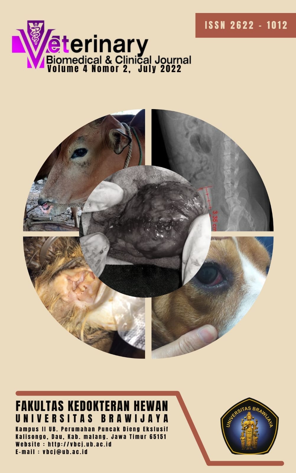
Urolithiasis is a disease caused by calculi, crystals, or excessive sediment in the urinary tract, which is generally composed of one or more types of minerals such as struvite, calcium oxalate, calcium phosphate, uric acid, and cystine. A 10-year-old female Pomeranian mix Pekingese dog was referred to Winadi Vet Animal Clinic Malang with clinical signs of dysuria, strangury, and hematuria. The physical examination findings were lethargy, caudal abdominal pain, and bladder distension. Radiological examination results indicated distension in the urinary bladder containing two stones occupying with clear margins and radiopaque-like opacity, with lengths of 3.44 cm and 3.35 cm, respectively. Cystotomy to remove the calculi was performed by incision on the midline.The urinary bladder was closed using a two-layer closure suture type, the first layer used simple interrupted, and the second layer used simple continuous suture with 3-0 polyglactin (Vicryl®) suture. The animal recovered on the fourteenth day after surgery
References
Antika, D. D., Kartanegara, A. A. S. and Rickyawan, N. 2021. Bladder Stones Removal Surgery Using Cystotomy in a Mix Breed Cat: A Case Report. Media Kedokteran Hewan, 32(3): 144 – 156.
Chew, D. J., and Dibirtola, S. P. 2004. Interpretation of Canine and Feline Urinalysis. Delawere: Nestle Purina.
Houston, D. M., Moore, A. E. P., Favrin, M. G., and Hoff, B. 2004. Canine urolithiasis: A look at Over 16,000 Urolith Submissions to the Canadian Veterinary Urolith Centre from February 1998 to April 2003. Can Vet J, 45: 225 – 230.
Kartika Y., Erina, N. And Asmilia. 2020. Blood Profile of Domestic Cats (Felis Domesticus) Suffering from Ear Mites. Jurnal Ilmiah Mahasiswa Veteriner (Jimvet), 5(1): 1 – 9.
Men, Y. V. and Arjentina, I. P. G. Y. 2018. Laporan Kasus : Urolithiasis in Rottweiller Mix Dog. Indonesia Medicus Veterinus, 7(3): 211 – 218.
Prakoeswa, F. R. S. 2020. Peranan Sel Limfosit dalam Imunologi : Artikel Review. Jurnal Sains dan Kesehatan, 2(4): 525 – 537.
Putri Z. U., Sulistiawati, E. and Sajuthi, D. 2018. Presentase Kejadian Hiperbilirubinemia pada Kucing. Proc.Of The 20th FAVA Congress & KIVNAS PDHI, Bali Nov 1-3:316 – 318.
Sardjana, I. K. W. and Kusumawati, D. 2011. Buku Ajar Bedah Veteriner. Surabaya: Airlangga University Press.
Tion, M. T., Dvorska, J. and Saganuwan, S. A.2015. A Review on Urolithiasis in Dogs and Cats. Bulgarian Journal of Veterinary Medicine, 18(1):1 – 18
Downloads
Published
How to Cite
Issue
Section
License
Copyright (c) 2022 Fenti Nursafitri, Dian Vidiastuti, Winda Syafitri

This work is licensed under a Creative Commons Attribution-NonCommercial 4.0 International License.