
The Routine Histopatological Examination-Chicken with Suspect Coryza (Stress Related Disease)
Keywords:
chicken, macroscopic, microscopic, histopathology, coryzaAbstract
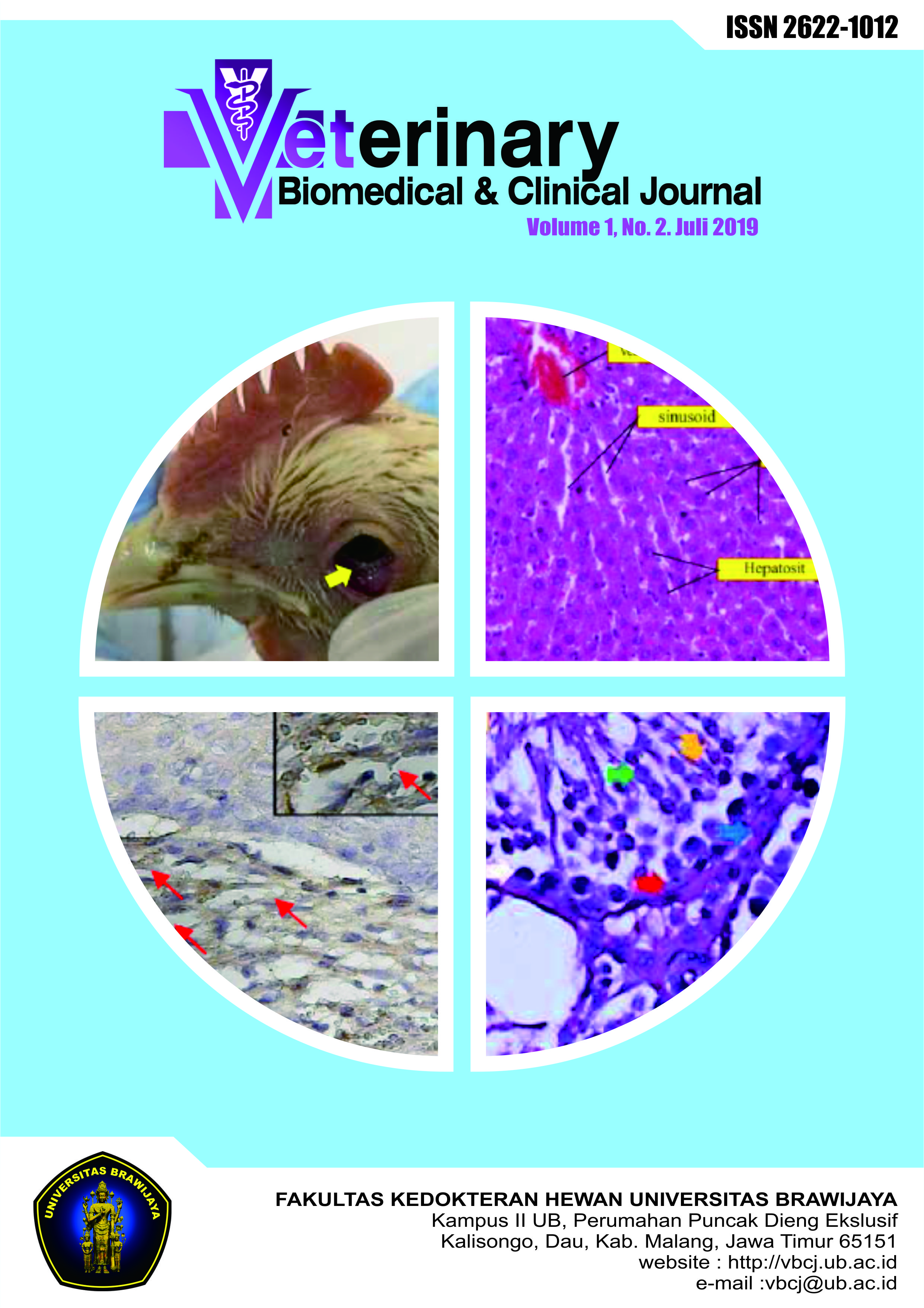
Coryza (snot) kown as respiratory disease in chickens caused by Haemophilus paragalinarum. Coryza is a disease that often occurs and results in large economic losses impact. The aim of this study was to look and diagnose changes in macroscopic and microscopic (histopathology) in chickens that have been examined and suspected of Coryza (snot) disease. The organs examined are palpabrae (eyes), trachea, pancreas and cerebellum. Examining and observing macroscopic changes, swelling of the palpabrae, tracheal hemorrhages and pancreatic hemorrhages and cerebellum were normal. Microscopic changes seen in palpabrae are oedema, hemorrhage and blood clot found, tracheal epithelial erosion, hemorrhage, multiplication of goblet cells and inflammatory infiltration of PMN cells, pancreas shown there is an inflammatory cell infiltration and fibrosis necrosis, and infiltrating cell inflammation in cerebellum. From examinations and observations it can be concluded that macroscopic and microscopic changes lead to the suspected Coryza (snot) disease.
References
Barlianto, W., M.S.C. Kusuma, S. Karyono, K. Mintaroem. 2009. The Development of Allergic Mouse Model Following Chronic Ovalbumin Exposure. Jurnal Kedokteran Brawijaya. 25(1).
Crispo, M., C.G. Senties-Cue, G.L. Cooper, G. Mountainspring, C. Corsiglia, A.A. Bickford, S.T. Stoute. 2018. Otitis and Meningoencephalitis Associated with Infectious Coryza (Avibacterium paragallinarum) in Commecial Broiler Chicken. J Vet Diagn Invest. 30(5):784-788.
Djojodibroto, D. 2007. Respirologi (Respiratory Medicine). Penerbit Buku Kedokteran ECG: Jakarta.
Garcia, J.I., I. Abdulkader, J. Larino-Novia, J. Forteza, J.E.Dominguez-Monoz. 2006. Histological Evaluation of Chronic Pancreatitis by Endoscopic Ultrasound-guided Fine Needle Biopsy. Gut. 55: 1661-1684.
Goljan, E.F. 2014. Rapid Review Pathology. Elsevier Health Sciences: USA.
Hinz, K.H. 1981. Serological Differentiation of Haemophilus paragalinarum Strains by their Heat Stable Antigens. In: Haemophilus, Pasteurella and Actinobacillus. M Kilian, W. Fredicksen and E.L. Biberstein (ed.). Academic Press. London. 1-10.
Mayer, J. and T.M. Donnelly. 2013. Clinical Veterinary Advisor, Bird and Exotic Pets. Elsevier: Missouri.
Miao, D., P. Zhang, Y. Gong, T. Yamaguci, Y. Iritani, and P.J. Blackall. 2000. The Development and Application of Blocking ELISA Kit for the Diagnosis of Infectious Coryza. Avian Pathol. 29: 219 – 225.
Nuryanto. 2012. Kajian Histopatologi dan Imunologi Ayam Pedaging yang divaksin Newcastle Disease Strain La Sota dan ditantang dengan Virus Newcastle Disease Strain Velogenik Indonesia (VND/Tasik/M13/2009). Institute Pertanian Bogor: Bogor.
Polanda, Agustin. 2004. Aneka Penyakit Ayam dan Cara Mengatasinya. PT Agro Media Pustaka: Jakarta.
Schimidt, R.E. and D.R. Reavill. 2014. Lesions of Avian Pancreas. Vet Clin Exot Animal. 17: 1-11.
Tabbu, C.R. 2000. Penyakit Ayam dan Penanggulangannya, Penyakit Bakterial, Mikal, dan Viral. Penerbit Kanisus: Yogyakarta.
Vazquez, C. E. Negrete-Abascal, S. Vaca. 2005. Haemophilus paragallinarum Secrets Metalloproteases. Canadian Journal of Microbiology. 51(10):893-6.
Downloads
Published
How to Cite
Issue
Section
License
Copyright (c) 2019 Albiruni Haryo, Janice Enola

This work is licensed under a Creative Commons Attribution-NonCommercial 4.0 International License.